VITREO RETINAL SERVICES & DIABETIC EYE CLINIC
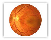
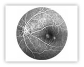
The Retina is like the film in a camera. It is the seeing tissue of the eye. When the focused light hits the retina, a picture is created and sent to the brain through the optic nerve (the nerve of the eye), thus giving us vision.
Retina has two parts: The Peripheral Retina and Central Macula. Macula being the central part, is capable of producing sharp and clear image. This clear images enable us to read , write and do all fine work. Conditions like Diabetes, Age related Macular degeneration and Macular holes can damage retina.
Retina Brochure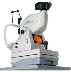
THE RETINAL EXAM INCLUDES :
A full inspection of the vitreous cavity and examination of the optic nerve, macula, retinal blood vessels and extreme peripheral retina. Once this is done, your doctor will make the decision whether or not to perform various retinal tests to further clarify the vitreous, macula, retina or choroidal diagnosis.
PROCEDURE FOR A RETINAL CHECK UP :
First through a routine check up of your refraction by a team of qualified optometrists. This will be followed by dilatation of your pupils for a detailed retinal exam by a consultant.
PRECAUTIONS FOR A RETINAL CHECK UP :
Following dilatation, it may be difficult to read or drive for several hours after the visit. If you have other systemic complaints ( diabetes, hypertension etc ), bring in your other medical records with details of the medications you are on.
- Vitreo-retinal problems
- Retinal Detachment
- Macular Degeneration.
- Diabetic Retinopathy
- Age-related macular degeneration
MEDICAL & SURGICAL RETINA DEPARTMENT :
Nirmals Eye Hospital is equipped with the most advanced equipments to diagnose and treat retinal diseases.
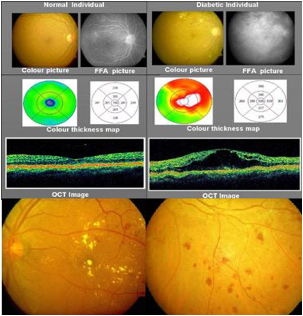
DIGITAL FUNDUS FLOUROSCIEN ANGIOGRAPHY :
A test to detect various Retinal conditions like Diabetic Retinopathy, Diseases of the Retinal Blood Vessels, Age Related Macular Degeneration, etc.
The digital imaging system gives us crystal clear images of the retina and aids in accurate diagnosis.
OPTICAL COHERANCE TOMOGRAPHY (OCT) :
OCT can measure retinal thickness precisely and repeatedly. It can measure minute changes, making it ideal for follow ups in retinal diseases, such as diabetes. Nirmals Eye Hospital is equipped with most advanceNidek Elite OCT system.
OTHER FACILITIES AT RETINA CLINIC :
Green (Argon) Laser for treatment of Diabetic Retinopathy, etc.
Nirmals Eye Hospital is one of the most advanced center for Retinal Surgeries like Retinal Detachments, Diabetic Retinopathy etc
SERVICES OFFERED :
Diabetic retinopathy diagnosis and management – Screening for diabetic retinopathy is important for individuals having diabetes of five years or more duration. It can destroy the vision silently over a period of time. Patients if diagnosed would need FFA and/or OCT to define the stage of the disease.
Advanced cases might need retinal laser treatment or vitrectomy surgery to restore the vision.
HYPERTENSIVE RETINOPATHY
Hypertensive retinopathy can affect the eye quietly or can present as loss of vision with sudden onset. Patients with hypertension needs to be examined from time to time and if evidence of retinopathy is found, they would need FFA, OCT, retinal laser or vitrectomy (surgery) depending on the kind of damage.
AGE RELATED RETINOPATHIES
Crucial areas of the retina can get affected during the old age. Generally the disease affects the people with age more than 50. Vision loss is generally slow and progressive. In few unfortunate patients there can be sudden acceleration of the disease process as well.
FFA and OCT are required to diagnose the extent of the disease.
Treatment options include medications, intravitreal injections, lasers, telescopic iol implantation and vitrectomy depending on the extent of injury.
RETINAL DETACHMENT MANAGEMENT
Retinal detachment generally happens in people with minus power glasses. It can also happen secondary to trauma, complicated surgery, other vascular retinopathies, some genetic disorders, pediatric retinopathies etc.
This is a surgical problem requiring scleral buckling or vitrectomy with/without gas or silicon oil injection.
CATARACT COMPLICATIONS MANAGEMENT
Retinal detachment generally happens in people with minus power glasses. It can also happen secondary to trauma, complicated surgery, other vascular retinopathies, some genetic disorders, pediatric retinopathies etc.
OCULAR TRAUMA
Injuries of the eye affecting the inside portion with affected vision or threatened affected vision requires surgical exploration and restoration of the normal anatomy.
PEDIATRIC RETINA
Do you see a white pearl your child’s eye? If yes, then contact us immediately. Most of the retinal disorders present in children like this. The condition can be as simple as bleeding at the back of the eye to serious disorders like tumours. Treatment depends on the problem child is facing. Premature child with low birth weight needs retinal evaluation early in the life to rule our ROP a vision threatening condition
ENDOPHTHALMITIS MANAGEMENT
The term represents a serious inflammation into the eye. Although there can be many causes leading to this problem most of the cases are secondary to some intraocular surgery. Patients require immediate intervention which may be in the form of multiple surgeries.
TREATMENT - MEDICAL :
- State of the art medical management of various retinal conditions
- Periocular injections – A simple injection given at the outermost layer of the eye ball to treat fluid collections at the crucial areas of the retina.
- Intraocular injections – The injection is given inside the eye. The procedure is done under all aseptic conditions under topical (drop) anesthesia.
- Retinal lasers – A 10 minute procedure done in OPD. Needs pupillary dilatation. Does not need any anesthesia. The defective areas on the retina are burned with the laser. Mainly used to treat vasular retinopathies.
- Laser for myopic retinopathies – A 10 minute procedure to delimit the retinal breaks to prevent a serious complication called retinal detachment. Needs pupillary dilatation. Does not need any anesthesia.
- Yag lasers for after cataracts – A two minute no anesthesia procedure to clean the artificial lens implanted in the eye during an earlier cataract surgery.
TREATMENT - SURGICAL :
VITRECTOMY :
- 20 guage – conventional method to perform a retinal surgery. Three holes are created in the eye to visualise and operate the retinal problem. The surgery is done under local anesthesia. Minmum duration of the surgery is 30 minutes. The patient might be implanted with gas or oil inside the eye. The oil needs to be removed later on. It is very important to maintain the position suggested by the doctor after the surgery.
- Surtureless transconjunctival – 23 and 25 guage retinal surgeries are the most modern sutureless methods to manage a retinal case. Post operative recovery is much faster than 20 gauge and patient is more comfortable as there are no sutures. The surgical time also reduces to a minimum of 20 minutes.
- Scleral buckling – A classical method to treat retinal detachments where a band or buckle is implanted around the eye to support the slipping retina. Minimum duation of surgery is 40 minutes and is performed under local anesthesia.
- Cryotherapy for retinal lesions – A 10 minute procedure performed under local anaesthesia. Here the early lesions on the retina are freezed with a cryopexy probe to prevent retinal detachment.
- Cyclodestructive procedure – Cryopexy probe is used to freeze the pressure maintaining tissue in the eye to control pressures in the eyes with uncontrolled gaucoma.
- Traumatic globe repairs – Surgical procedure depends on the kind of presentation the patient has. The management can range from simple suturing to complicated combined procedures like combination of scleral buckling with vitrectomy.
- TUMOR EXCISION - This procedure is done for the patients with tumors threatening the vision or life. The procedure can be followed up with chemotherapy or radiotherapy.